|
| Molecular profiling of Embryonic Stem Cells and Embryonic Carcinoma Cells:
Embryonic stem cells (ESCs) and embryonic carcinoma cells (ECCs) are closely related yet developmentally distinct cell types. ESCs are derived from the inner cell mass of blastocyst-staged embryos and ECCs; the stem cells of teratocarcinomas (mixed germ cell tumors) are derived from progenitors of the germline. Both cell types share the general properties of pluripotent stem cells with self-renewing potential and can give rise to derivatives of all three embryonic germ layers. Pluripotent stem cells can potentially provide sufficient numbers of differentiated cells to treat a wide variety of human conditions, including heart disease, diabetes, and many neurological disorders. However, several hurdles exist concerning the safe use of these cells. These cells could be genetically unstable and cause aberrant changes during propagation. ECCs, though are clinically useful, are malignant counterpart of ESCs with oncogenic potential. Identifying factors that turn on the oncogenic state versus those that enhance proliferation and self-renewal is critical for their clinical use. In this study, a comprehensive quantitative proteomic analysis was carried out to identify the differences between ESCs and ECCs that contributes to their pluripotency and malignancy. Some of the key molecules that were significantly higher in ESCs include Beta-galactoside-binding lectin (LGALS1), undifferentiated embryonic cell transcription factor-1 (UTF1), DNA cytosine methyltransferase 3 isoform-B (DNMT3B), melanoma antigen family-A4 (MAGEA4), and interferon induced transmembrane protein-1 (IFITM1). CD99-antigen (CD99), growth differentiation factor-3 (GDF3), cellular retinoic acid binding protein-2 (CRABP2), and developmental pluripotency associated-4 (DPPA4) were among the ones significantly higher in ECCs. Through this study, Isotope labeling based quantitative proteomic approach was shown to be a powerful method to identify differences between cells originating from different genetic backgrounds.
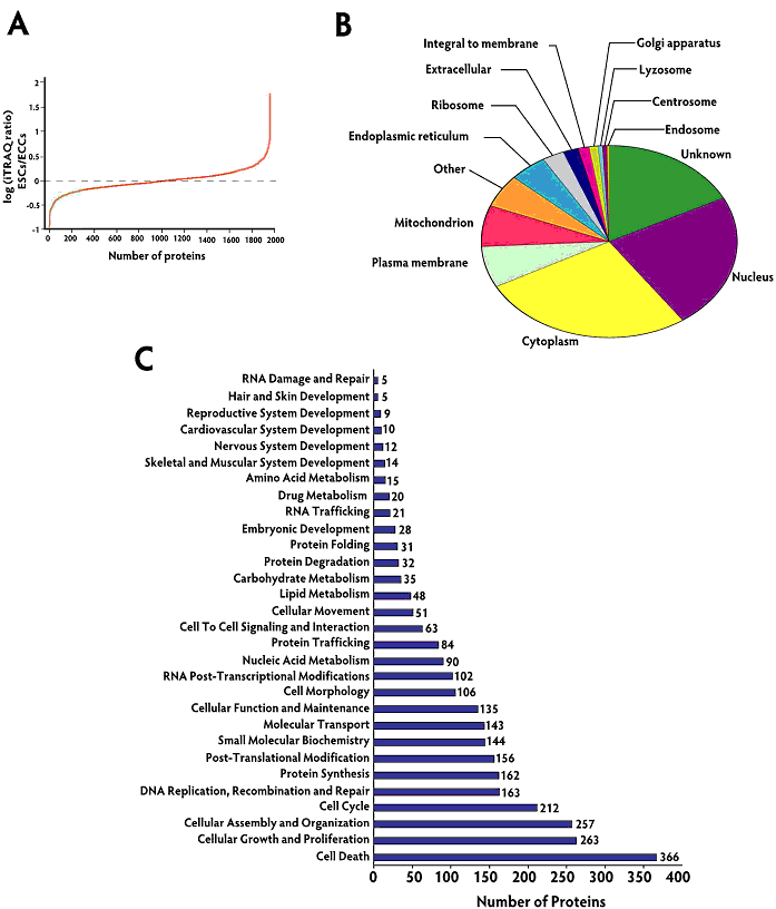
Localization and functional annotation of proteins identified from ESCs and ECCs. Panel A shows the distribution of iTRAQ fold changes (proteins expression levels) observed between ESCs and ECCs. Panel B shows the gene ontology analysis for cellular localization of all the proteins identified. Primary and alternate localization data was downloaded from human protein reference database (www.hprd.org) and Panel C shows functional classification of all the proteins quantitated in this study. Using Ingenuity pathway analysis tool, proteins justifying specific biological function significantly (p <0.05) are listed. |